All About Angiogram

Overview: Angiogram
A coronary angiogram is a procedure that uses X-ray imaging to see your heart's blood vessels. Coronary angiograms are part of a general group of procedures known as heart (cardiac) catheterizations. Cardiac catheterization procedures can both diagnose and treat heart and blood vessel conditions. A coronary angiogram, which can help diagnose heart conditions, is the most common type of cardiac catheterization procedure.
During a coronary angiogram, a type of dye that's visible by an X-ray machine is injected into the blood vessels of your heart. The X-ray machine rapidly takes a series of images (angiograms), offering a look at your blood vessels.
A slim catheter (a flimsy, empty plastic cylinder) is strung through the biggest supply route in your body (the aorta) by means of the wrist or the groin artery until it arrives at the coronary artery of the heart. An extraordinary x-ray beam delicate color (contrast) is infused and dynamic x-rays are taken of the veins as the differentiation travels through them.
Why is it done?
The test is generally done to see if there's a restriction in blood flow going to the heart.
Your doctor may recommend that you have a coronary angiogram if you have:
- Chest pain (angina)
- Ache in your chest, jaw, neck or arm that can't be explained by other tests
- New or increasing chest pain (unstable angina)
- A heart defect you were born with (congenital heart disease)
- Abnormal results on a noninvasive heart stress test
- Other blood vessel ailments or a chest injury
- A heart valve problem that needs surgery
How does an angiogram work?
A small amount of a radioactive tracer emits x-rays of the bloodstream. The tracer “tags” red blood cells, making it possible to gather images of blood circulation in the heart. X-rays will detect the traces. After the x-rays are converted into an electrical signal, they go to a computer, which creates an image of the chambers of the heart.
How can you prepare for the angiogram test?
In few cases, coronary angiograms are performed on an emergency basis. Mostly, though, they're scheduled in advance, giving you time to prepare.
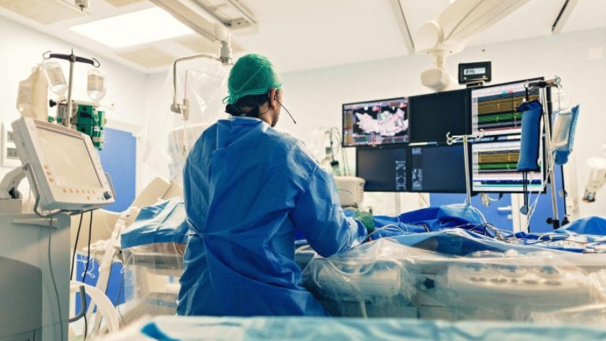
The Angiograms are performed in the catheterization (cath) lab of a hospital. The hospital team will give you specific instructions and talk to you about any medicines you take. General guidelines include:
- Don't eat or drink anything after midnight before your angiogram.
- Plenty of clear fluids up to 3 hours prior to a booked angiogram.
- Take all your medications to the hospital with you in their original bottles. Ask your doctor about whether to take your usual morning medications.
- If you have diabetes, ask your doctor if you should take insulin or other oral medications before your angiogram.
Medications:
- Please acquire every one of your meds with their unique packaging. You might have to stop, start, or change a portion of your medications before the angiogram procedure starts.
- You will get a letter with significant directions about taking your meds, also, your arrangement date. Kindly read this data cautiously.
General:
- On the off chance that you get directions to shave the groin area, adhere to these guidelines. On the off chance that you do not get directions to shave the area, it implies the medical caretaker will do it for you when you go to the hospital.
Valuables:
- Do not bring cash, valuables, or a lot of personal items and clothing
- Remove all jewellery and nail polish. You may keep your glasses, hearing aid (s), and denture(s) on during the procedure.
- Wear loose-fitting clothes and flat shoes.
Risk factors: Angiogram
An Angiogram is not performed in the accompanying cases:
- A critical history of an unfavourably susceptible response to differentiate color
- Pregnancy
- Presence of bleeding disorders
- Presence of kidney disorders
- Using blood-thinning prescriptions
What happens during an Angiogram procedure?
You will go to the catheterization lab (cath lab) for the angiogram.
- You will lie on the x-rays table. The Nurse will interface you to a heart monitor.
- The nurse will clean the area picked for the angiogram with a cleaning arrangement. Once it is cleared it's recommended to try not to contact this area.
- The nurse will put a sterile (without germ) wrap over you to keep the area clean.
- The cardiologist will inject a local sedative (freezing) into the groin area or wrist area part (radial artery).
- Once the area is frozen, the cardiologist will insert a sheath (like a large IV) into the femoral artery (found in the groin area) or radial artery.
- Through this sheath, the cardiologist will direct little catheters (wires) into the coronary arteries.
- Small measures of colored dye will be injected through these catheters to see the coronary arteries. It is not unexpected to feel a warm sensation for the moment.
- Patient need be ready to pause your breathing and give a deep cough off chance that the cardiologist asks you.
- It is typical to feel some gentle inconveniences during the angiogram. Notwithstanding, tell the cardiologist on the off chance that you are not comfortable with pain.
- Once enough photos are captured of your coronary (heart) arteries, the cardiologist will eliminate the guide wires however, leave the sheath in. Then he will look into the angiogram results.
- You will get back to the recuperation area.
What happens after the test?
Postoperatively after Angiogram:
- Plenty of rest is advised
- Drink plenty of liquids
- The wound area should be clean and dry
- Observe the wound for any signs of infection like drainage from the wound, swelling, redness, pain, and fever
- Avoid showering, tub baths, or swimming until the incision site is cleaned completely
Results of aniogram
Normal angiogram or mild narrowing (blockages)
Now and again, the angiogram can be ordinary with no narrowing. In different cases, the angiogram might show mild narrowing, with under 50% narrowing of the artery diameter. This is uplifting news. Your doctor will talk about the outcomes with you and you should circle back to your referring physician.
Blockages in little arteries
It is possible to have narrowed in small veins or branches providing just small spaces of the heart. These are generally left alone if the diameter of the arteries is below 2 mm. For the most part, it isn't advisable to put stents in tiny arteries. Angiogram treatment with normal heart medications is ordinarily suggested in these cases.
Serious blockage in a couple of significant arteries
In situations, where a couple of arteries have extreme narrowing, your primary care doctor might suggest an angioplasty procedure. This system is typically done simultaneously as the angiogram, permitting the specialist to open up any narrowing with a special device called a balloon or angio stent.
Serious blockages in numerous supply routes
At times, we see numerous arterial routes with serious blockages. This is more common in patients with diabetes. Cases with several blockages are regularly treated with sidestep bypass surgery. The angiogram is halted as of now to permit time for conversation with you, your family, your alluding specialist doctor, and a cardiovascular specialist surgeon.
How much does angiography cost in Gleneagles hospital?
The rates as per heart angiography cost in Mumbai:
Investigation
Approximate cost (In INR)
ECG
370
2D ECHO
3040
TMT (Treadmill Test)
2890
Chest X-ray
640
Day Care Cath Coronary Angiogram
CT Coronary Angiogram
Note:
The above-mentioned prices are approximate prices of those procedures respectively.
You can make an appointment or call on 91-022- 67670101 on the website to know angiogram cost in Mumbai.
Why Gleneagles Hospital for Angiogram?
The Cardiac Science team at Gleneagles Hospital, Parel, Mumbai comes with an experience of over 20 years. The Cath- lab here, is with integrated Intravascular Ultra sound(IVUS) and Fractional Flow reserve(FFR) which is an advanced technique. IVUS can be used to assess vessel/lumen diameter, lesion length, help determine the amount of plaque build-up in a vessel and its composition. This aide in getting accurate reports, thereby planning of the required line of treatment. Further this optimizes the results post angiography as it helps to check if the stents have been properly placed and fully deployed. It can also help measure the effectiveness of balloon angioplasty or stenting during follow ups. The benefit of advanced technology is it saves unnecessary coronary interventions and patients could be managed with medications alone. Gleneagles hospital, Parel, Mumbai is known for its honest opinion and zero unwanted cardiac interventions.


